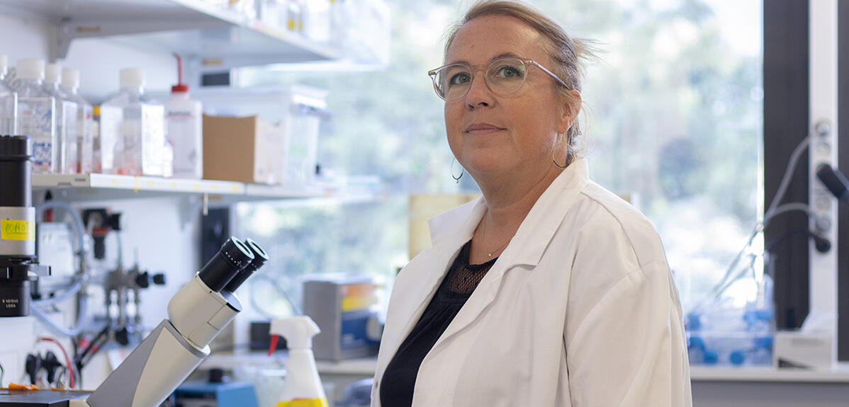
With a background in biology and chemistry, Stéphanie Descroix works in a highly multidisciplinary research field: microfluidics. Using this technology, he creates mini-organs on a chip. Tools that open huge perspectives, especially in oncology…
In a conversation with Stéphanie Descroix, CNRS research director and team leader at the Institut Curie in Paris, one character trait stands out: her “positive attitude”. Let’s judge: his workplace? ” It is a beautiful center, the most beautiful place for my research “, she says. His work? He is ” super », “ super satisfying “. His career? ” I was very lucky! » And his associates? Many are from ” great colleagues “. ” She creates such a good atmosphere in her group that it is hard to leave. », notes Charlotte Bouquerel, who worked with her for four years, as part of her doctoral internship.
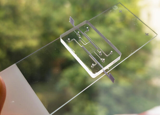
A microfluidic chip on which the organoid will grow.
But the researcher also excels with her research… at the cutting edge of technology! This is because his group, the “Macromolecules and Microsystems in Biology and Medicine” team, is one of the world leaders in a recent field that promises a revolution in the understanding of human physiology and pathologies and their management: organs on chips.
Organs on a chip are new technologies designed to reproduce certain cellular, biochemical, physical and physiological characteristics of human organs and tissues.
Also say ” organs on a chip » (from their English name), « organs on a chip are new technologies designed to reproduce certain cellular, biochemical, physical and physiological characteristics of human organs and tissues, such as their three-dimensional structure, their physico-chemical environment (oxygen level, acidity, etc.) or their functions », explains Stéphanie Descroix. These systems are produced using microfluidics, a technology born thirty years ago that is booming today.
At the crossroads of biology, physics, chemistry and engineering, microfluidics enables the production of miniature devices on small chips of glass, silicon or plastic. Small in size (a few square centimeters), these platforms contain a set of etched or patterned microchannels, linked together to perform a specific function such as mixing components or controlling the biochemical environment.
The art of doing better with less
In practice, ” organs on a chip are obtained from cells and molecules of the extracellular matrix, the “cement” that holds cells of the same tissue together. The assembly is injected into a microfluidic chip where it self-organizes to acquire a three-dimensional structure that can be similar to that of a real organ. », explains the head of the research.
The advantages of organs on a chip are huge! First of all, microfluidics enables the control of various biological, physical or physicochemical parameters: cell composition, extracellular matrix, oxygen level, acidity, applied forces, etc.; which makes it possible to approximate the characteristics and conditions inside real organs or tissues. As a result, organs-on-a-chip should be more reliable experimental tools than simple cells in culture in the future.
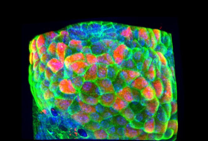
Fluorescent microscopic image of intestinal villi on a microfluidic chip.
Then, they make it possible to carry out numerous experiments with very little biological material: ” tens of thousands of cells for an organ on a chip, which can represent a few square millimeters of the original organ “. Finally, compared to animal or human tests, they allow for faster and lower cost. In short, as Stéphanie Descroix points out in the book’s chapter on microfluidics, these systems allow ” do better with less » !
Multidisciplinarity anchored to the body
How does a researcher specialize in such a specialized field? In fact, this resident of Ile-de-France born in Fontenay-sous-Bois (94) has always been immersed in an environment conducive to scientific curiosity: “ My parents, both scientists but in industry, had a real appetite for science, which they passed on to my brother – now a maths teacher – and me. After that I married a researcher and now I have two children who also love science. “, she confides.
She also showed a strong taste for different disciplines very early on: in high school i liked math, biology, history, german and also physics-chemistry “. But when graduation is in hand, she has to make a choice.
In the future, organs on a chip should be more reliable experimental tools than simple cultured cells.
She then enrolled in biology at the University of Natural Sciences in Créteil (94). As soon as she mastered the science and techniques of biochemical and biological engineering, she was overtaken by the need for multidisciplinarity. she ” it forks » then towards a diploma of advanced studies, this time specialized in analytical chemistry (this part of chemistry devoted to the analysis of chemical products), at the Pierre-et-Marie-Curie University, now the Sorbonne University.
After his thesis was obtained in 2002, the one that says ” often in a hurry, to the point that (his) tennis teacher keeps telling (him) to do three or four rallies before going to the net and finishing the attack! », bypasses the traditional post-doctoral internship, which normally completes the training of researchers, and decides on a position at the University of Orsay as a temporary teaching and research associate. But very soon, she feels ” more (not) a place in research than in teaching », and tried to pass the CNRS entrance exam, which she won in 2004.
Putting cancer on a chip to move towards personalized medicine
Microfluidics? Stéphanie Descroix took her first steps there after joining the CNRS: “ In that timeshe says, this technology began to take off significantly in France. Also, I wanted to with my host team at the time – Physical-Chemical Laboratory for Electrolytes, Colloids and Analytical Sciences. –combine with bioanalytical approaches (which allow quantitative measurement of a biological object, editor’s note).” However, she notes, ” It took me a long time to become an expert in microfluidics and organs-on-a-chip…and my learning is far from over! »
It took me a long time to become an expert in microfluidics… and my learning is far from over!
Now, at the Curie Institute, which she joined in 2011, the researcher and her colleagues are developing specific organs on a chip: ” patients with tumors on a chip “. As she explains, ” These are micro-tumors created from different cells of the same patient: cancer cells, but also others naturally present in tumors, such as immune cells and blood vessel cells. “.
Thanks to this type of tool, the researcher hopes to realize a big dream: to develop personalized medical systems that would allow testing a patient’s response to chemotherapy or immunotherapy (two types of cancer treatment); these therapies can be more or less effective depending on the characteristics – especially the genetic ones – of each tumor. ” If we succeed in developing such tools, they could help deliver the most effective therapy directly to the patient. Which would increase his chances of survival », hopes Stéphanie Descroix.
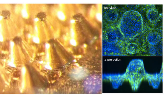
On the left, a brass mold imitating the structure of a gut. It is used to shape a 3D collagen structure to create a gut-on-a-chip. The right side shows the intestine on a chip with cells that have colonized the structure.
During a recent study, the researcher and her colleagues demonstrated the feasibility of this concept on about ten patients. Now it remains to repeat the study on a larger scale. do this, a large clinical trial with around two hundred patients is planned. It is expected to be launched in the next six months.
Many discoveries in perspective
But it’s not just applied research! ” My team also conducts basic research to improve our knowledge of organs and their diseases. The possibility of working on those two sides of research that we tend to oppose, although they feed each other, is a specific thing that interests me. », points out Stéphanie Descroix, with a certain pride.
In this area, the researcher “ To have fun » in particular to try to answer a few very specific questions concerning the intestines, ” an excellent organ, but too often underestimated “. For example, during recent work published specifically with Danijela Vignjević, a cell biologist at the Institut Curie, she co-developed a gut-on-a-chip that allowed us to learn more about the establishment of the different types of cells that make up the intestinal epithelium, the tissue that covers the inner lining of the thin bowels. We wanted to know what drives the spatial organization of these different cell types, knowing that they are not located randomly, but at specific levels in the epithelium. “, she explains.
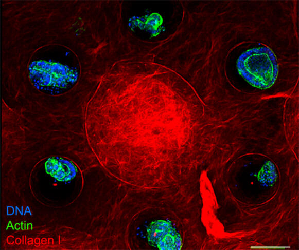
Fluorescence microscope image of a villus surrounded by a crypt, an intestinal organoid is deposited in each crypt.
And bingo! The team leader and her colleagues showed that although the specific geometry of the epithelium in the crypts (cavities) and villi (folds) partially regulates the position of the cells in this tissue, this is not enough. It also requires the presence of specific cells, called fibroblasts, which produce substances (growth factors and collagen, the main component of the extracellular matrix) essential for the correct position of the cells. In fact, concludes Stéphanie Descroix, “ the potential of organs on a chip is huge. In the years to come, they should lead to many discoveries. Both in basic and applied research and in the clinic ! “. ♦
Read on our pages
THE organoids: from mini-organ to maxi-power
